
Unit IV-Circulatory System
Chapter 11
Anatomy and Physiology of the Arteries
1. General Remarks:
On leaving the right ventricle
blood goes to the pulmonary artery. From the left ventricle, blood goes to the
aorta; from the aorta
there are branches to all parts of the body. The pressures in the pulmonary
artery are about one-fifth those in the aorta and its branches;
but the structure of the pulmonary artery is like that of arteries anywhere. The
pulmonary circulation will be considered later
(Chapter 15).
2. Structure of the Arteries:
The blood in the arteries is at
higher pressure than in the corresponding veins. For reasons
which are not understood, the arterial wall becomes thicker than the venous
wall. This thickening is usually achieved by
thickening of the middle layer with muscle, as is the case in the smaller
arteries. Sometimes, particularly in the aorta, the middle layer is thickened
with elastic fibers. Arteries in which muscular thickening
predominates are called muscular arteries; the others are called
elastic.
|
|
|
|
Figure 226 shows these differences. Part (a) of this figure shows a small muscular artery and (b) shows the corresponding vein. Figure 227 (a) is thoracic cross section through the aorta, and (b) is a cross section through a large vein (inferior vena cava). In the arterial cross sections note that there are three major layers of tissue. The innermost, called the endothelium, is very smooth and very thin. Its smoothness is important because blood tends to clot when it is in contact with rough surfaces. The middle layer, here shown as muscular, stretches easily when it is distended by the pulse of blood. During diastole, it recoils, and the blood runs off. The outermost layer is a protective layer of connective tissue.
It may be noted that the muscular coat usually contracts in the absence of a blood pressure to keep it open. Thus, in a freshly dead animal, the arteries do not seem to contain any blood at all. The Greeks, noting the absence of blood, assumed that they were channels for the delivery of air. In fact the word artery really means air tunnel.
3. Distribution of the Arteries:
The blood which lçaves the left ventricle goes into the aorta; from the aorta branches to the arteries lead to all the major parts of the body. The very first branches of the aorta are the coronary arteries, which supply the heart muscle. As the aorta arches over and behind the heart it gives rise first to the brachiocephalic artery, also known as the innominate artery, which branches immediately to form the right subclavian and the right carotid. The next branch of the arch is the left carotid; then the left subclavian takes its origin from the arch.
Within the chest the aorta gives rise to a number of small branches. These supply the intercostal muscle and the diaphragm. There are also branches to the esophagus, the pericardium and the lungs (bronchial arteries).
The first major branch of the aorta which occurs after it has passed through the diaphragm is the celiac artery. This supplies the part of the stomach, the spleen, and the liver. Arterial blood to all the remaining digestive organs, except the last parts of the colon, is delivered through a branch just below the celiac artery; this is called the superior mesenteric artery. Two large arteries now stem from the aorta, at right angles to it; they supply the kidneys (renal arteries). Small branches to the abdominal wall are also shown. Just before it breaks up into the iliac arteries, there is a small branch which supplies the transverse and descending colon. It is called the inferior mesenteric artery. Finally the aorta divides into the two common iliac arteries. These divide again, forming the internal and external iliac arteries. The internal iliac artery supplies the organs of the pelvis; the external iliac artery supplies the lower extremity.
The details of the arterial distribution are shown in Figures 228 to 233. Figure 228 shows the relationship of the aorta to the heart, the distribution of the coronary arteries, and the major branches of the aortic arch. Figure 229 shows the branches of the descending aorta within the chest. Figure 230 shows the relationships of the abdominal aorta and its major intra-abdominal branches. Figure 231 shows the arterial supply of the head and neck. The arterial supply of the arm is shown in Figure 232; the arteries of the leg and thigh are shown in Figure 233.
4. Arterial Pressure and the Pulse:
As has been noted, the heart is an intermittently acting pump; the blood from it is not ejected into the arterial system steadily but rather in a pulse. With each ventricular systole the amount of blood in the arteries increases and the pressure within the arteries rises. During diastole the blood which was ejected by the ventricular systole runs off from the arteries into their most distant branches. The structure of the arteries discussed in Part 2 favors this function.
The pulse wave travels a good deal faster than the ejected blood itself. This is due to the fact that the arteries, when stretched, recoil, transmitting a pressure wave through their walls along the length of the arteries. The movement of the blood follows this pressure wave. The arterial pulse usually moves very fast (about 5 m/s) so that it reaches the furthermost portion of the arterial tree almost immediately after systole of the ventricles is over. The passage of blood is much slower although still very fast. For example, the time for blood to go from the aorta to the arteries of the tongue is only 2 or 3 seconds.
The pressure which is in the arteries is spoken of as the arterial blood pressure; the customary unit for the description of arterial blood pressure is in mm Hg (millimeters of mercury). A pressure of 100 mm Hg means that the pressure will displace a column of Hg in a U tube so that the highest part of the column is 100 mm above the lower part of the column. One can get a better picture of this by expressing pressure in terms of meters of blood. 100 mm Hg corresponds to approximately 1.3 meters of blood, so that if the pressure in an artery were 100 mm Hg and if the artery were connected to a long tube open to the outside, blood would rise in it 1.3 meters. This was, in fact, precisely the way in which arterial pressure was first measured by an English clergyman named Stephan Hales who connected a tube to the femoral artery of a mare without anesthesia. In this experiment the blood rose 8 ft 3 in (almost 2. 5 meters) in the tube indicating that the animal had a rather high blood pressure.
Devices which measure the pressure of the arterial blood are called manometers. Of these, the one most commonly used in medical practice is called a sphygomomanometer, which operates on the principle that when the pressure in a cuff around an artery exceeds the pressure in the artery, the artery will collapse and the pulse will disappear. This will be considered in more detail later in this chapter.
As was noted before, the pressure in the arterial system varies continuously between systole and diastole. The highest pressure attained is called the systolic pressure. During diastole the pressure falls to its lowest level, the diastolic pressure. Their difference corresponds to the amount of the arterial pulsation, is called the pulse pressure. Thus, if the systolic pressure is 120 mm Hg and the diastolic pressure 80 mm Hg, the pulse pressure is 40 mm Hg.
The mean arterial pressure lies between the systolic and the diastolic pressure. It is sometimes convenient simply to average the two values, but a more accurate method of obtaining this mean value is to give the diastolic pressure twice the representation of the systolic. Thus, if the systolic pressure is 120 mm Hg and the diastolic is 80 mm Hg, we could estimate the mean pressure to be 100 mm Hg (the average of the two values) or more accurately we could describe it as:
| 120 + 80 + 80 | |
| = 93 mm Hg | |
| 3 |
There is considerable confusion, especially among the public, about what is meant by arterial blood pressure. Most people tend to think that their blood pressure is best described as the systolic pressure and one often hears people saying, "My blood pressure was 160," when the real statement is "My systolic blood pressure was 160 and my diastolic was 60." In the absence of further information one might take the value of 160 as indicative of high blood pressure but when the diastolic pressure value is also given it is clear that the mean blood pressure is still 93 ( [160 + 60 + 60] / 3 = 93) and the mean blood pressure is not elevated at all.
5. Measurement of Arterial Pressures:
The obvious method for measuring blood pressure-connecting the artery to a vertical glass tube and observing the height to which the blood rises-is neither practical or accurate. It is impractical because it requires making an opening into the artery. It is inaccurate because the rapid variations in arterial pressure with each pulse beat cannot be followed by the heavy columns of blood.
Better methods involve the use of devices which measure pressures by the displacement of very small objects, whose movement is recorded electrically. There are many such devices, but all suffer from the disadvantage that the moving object must be in direct contact with the blood inside the artery, so that the artery has to be punctured.
The most practical method for measuring the blood pressure is by the use of an inflatable cuff placed around the arm. When air is introduced into such a cuff the pressure inside it can be measured directly. When this pressure is higher than the systolic arterial pressure, the artery is compressed shut between the cuff and the bone. This results in the disappearance of the pulse at the wrist.
When the pressure in the cuff is lowered to a point just below the systolic pressure, the pulse reappears. The artery, however, is not open all the time. It opens when the blood pressure is higher than the cuff pressure and closes when it falls below cuff pressure. The movements of the artery give rise to a sound, which can be heard best over the artery just below the cuff-that is, on the inside of the elbow.
When the cuff pressure is just above diastolic pressure, the artery opens and closes just as before, though it is open longer. When, however, the cuff pressure is below diastolic pressure, the artery never closes and the sound disappears.
In order to measure the arterial blood pressure, the observer places the cuff around the upper arm, and increases cuff pressure until the pulse disappears at the wrist. The cuff pressure is lowered slowly, while the observer listens, with a stethescope over the artery, just below the cuff. When the cuff pressure is higher than the systolic arterial pressure, no sounds will be heard. As cuff pressure falls below systolic pressure, the artery begins to generate sounds. The cuff pressure at which these sounds first appear is a good measure of systolic pressure.
Further lowering of cuff pressure results in a series of changes of the sounds heard. These are called Korotkow sounds, and they are not completely understood.
The cuff pressure at which the sounds just disappear entirely corresponds quite closely to the diastolic pressure as measured by other means. This method of measuring arterial blood pressure is called the "indirect" method and is the one used mostly in man. There is little argument about the accuracy of the systolic pressure measurement by this method. Some authorities, however, believe that the diastolic pressure is measured best before the sound disappears, while others prefer total disappearance. Which of these should be used is a matter for individual and institutional preference.
6. Normal Values of Arterial Pressure:
It is usually the practice to
record arterial pressure by writing the systolic pressure first, then a stroke,
then the diastolic pressure.
A person with a systolic pressure of 120 and a diastolic pressure of 80 would
thus be said to have blood pressure of 120/80. The normal values
of arterial pressure are very variable. In young people the systolic pressure
may be anywhere between 100-140 while the diastolic pressure may
be anywhere between 60-90. The blood pressure tends to increase somewhat with
age. The following table shows the blood pressure which are
considered questionably high in recent life insurance statistics.
Blood pressures lower than these
are not necessarily normal, nor are blood pressures higher than these
necessarily indicative of high blood
pressure. Only a physician who has further information regarding the history and
physical condition of the patient can make an intelligent
decision about the meaning of the patient's blood pressure.
7. Regulation of Arterial Pressure:
The body controls mean arterial
pressure better than systolic or diastolic pressures. This control is
accomplished through adjustment of
the peripheral resistance to the cardiac output. During activity, the
active tissues increase the return of blood to the heart and
thereby increase the cardiac output. If the arteriolar resistance did not
change, the increased flow would result in changes in arterial
pressure. For example, when the cardiac output is six liters per minute, the
mean arterial pressure is about 90 mm Hg. In exercise, the
cardiac output may increase four times, but the mean arterial pressure changes
only slightly. This means that the resistance of the arterioles
must have decreased more than four fold. In the opposite direction, blood loss
often reduces the cardiac output, but usually the blood
pressure is not affected as much as the cardiac output, because arteriolar
resistance increases.
Some of the factors which
influence the resistance of the arterioles are local; these will be taken up in
the next chapter.
The barostatic reflexes (aortic arch and carotid sinus), already
considered from the standpoint of heart rate control, are extremely
important in regulation of the arterial blood pressure. When blood pressure
rises, the arterioles dilate. Their resistance decreases and the
blood pressure falls. Falls in blood pressure have the opposite effect. The
overall action of the barostatic reflex is to keep
the mean blood pressure constant within a rather narrow range.
For some
reason, not yet well understood, the barostatic reflexes do not operate
normally in persons with high blood pressure. The mean
blood pressure may be increased 50% to, say, 140 mm Hg. This is due to general
arteriolar constriction. Yet the barostatic reflexes do not
operate to dilate the constricted arterioles. It is not known whether their
failure results from disorders in the receptors or the centers.
Animal experiments suggest that the receptors are at fault. Thus, in dogs with
high blood pressure, the sensory nerves of the baroreceptor
reflexes discharge at the same rate as if the blood pressure were normal. When
the blood pressure of such animals rises above the accustomed
(high) level, the receptors discharge at an increased rate; and when it falls
below the accustomed level, they discharge at a decreased rate.
In other words, the receptors behave in a normal way, but instead of operating
to conserve the normal blood pressure level, they act to
conserve a high one. To make an analogy with a thermostat, it is as if the
temperature scale had been moved in a home thermostat. When one
sets the temperature to 70 on the scale, the real setting might be at 90. The
heating and cooling systems would still be operative but instead
of holding a desired temperature of 70, they would work around the erroneously
set temperature of 90. How this type of error comes about in
animals in blood pressure regulation is completely unknown, but it seems certain
that it occurs in humans with high blood pressure. High
blood pressure will be considered in further detail in
Chapter 16.
8. Abnormalities in Arterial Circulation:
Abnormal pulse pressures are
brought about in a variety of ways. The most dramatic is that which occurs in
complete insufficiency of the
aortic valves. When the heart has completed its systole the blood simply falls
back into the relaxed ventricle and the diastolic pressure
consequently becomes zero. In the next systole of the heart the blood which fell
back into the ventricle is ejected as well as the blood which
was returned to the left ventricle from the pulmonary veins. This means that the
heart must eject a much larger volume of blood into the
arteries per stroke than is normal. This large volume produces a greater
stretching of the arterial system and consequently a high systolic
pressure. In such a case, the systolic pressure might be 200 while the diastolic
was 0. The mean pressure is only 67 ( [200 + 0 + 0] / 3) which
is obviously not abnormally high. The pulse pressure in such a person would be
very high corresponding to the very large stroke volume and
sometimes the diagnosis of aortic insufficiency can be made simply by feeling
the pulse in the radial artery.
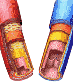
Another condition which produces
abnormalities in the arterial pulse is arteriosclerosis (hardening of the
arteries). When this occurs the
blood, ejected by the ventricle during systole, is ejected into a stiff arterial
system and the pressure rise which occurs during systole is
much greater than normal. This is usually seen in older people where a systolic
pressure of 180 may go along with a diastolic pressure of 50.
This should not be taken to indicate high blood pressure, in the true sense of
the word, but only shows arterial stiffness.
Athletes sometimes have a very
slow heart rate. The cardiac output per minute is usually normal, but since the
heart rate is slow, the amount of blood ejected per stroke is unusually large.
This results in over-distension of the arteries during systole, but the diastolic
pressure is low since there is a longer time during which the blood can leave
the arterial system before the next systole.
There are many other conditions
which can produce abnormal pressures in the arterial system such as aortic
stenosis, a rapid heart rate, an
increased cardiac output, and so forth. The student should try to reason out the
effect of these changes on arterial pressures.
The obstruction of small arteries
in most organs probably occurs much more often than is recognized. The probable
cause of such obstructions usually thrombus formation upstream. If the thrombus
is dislodged and moves down-stream, it will interrupt the blood supply in the
downstream area which it obstructs. Oddly enough, this is not usually a serious
matter, for in most parts of the body there is a potential supply of
arterial blood vessels which can reach the same part by way of other channels.
When an arterial channel is obstructed, other channels communicate
with it past the obstruction, and will in effect reestablish the circulation. An
extreme example of this is seen in a congenital defect
coarctation of the aorta. The aorta is severely constricted, usually in
the chest; sometimes so severely that no flow at all can occur to
the abdominal aorta via the normal passageway. This can be entirely symptomless.
The branches of the aorta which do occur in the chest make
branches which communicate with the branches of the abdominal aorta. Thus, flow
to the abdominal aorta and its branches is by a roundabout pathway.
Collaterals have taken over the function of the main vessel.
The factors
which influence the growth and development of collaterals are not well understood.
It seems probable that collaterals are developed in any part of the body whose
activity is sometimes greater than its normal blood supply. For example,
well developed leg muscles probably have good
collateral blood supply as well as their normal blood supply. There are certain
areas of the body (notably the myocardium and the brain) where
collaterals are usually so slow to develop after an arterial obstruction that
they come too late to preserve the life of the tissue whose arterial
blood supply has been obstructed. There has been evidence recently which
suggests that collateral circulation in the myocardium may be developed
by exercise. Persons with a history of regular exercise appear to have as many
episodes of coronary arterial obstruction as others, but they die
of them only half as often. Presumably, this is a consequence of the fact that
their exercise has promoted collateral development in the myocardium,
so that the area which has lost its own blood supply can receive enough blood by
other routes to maintain it alive.
At the present time, there is no
known method for increasing the collateral blood supply of the brain. Arterial
obstruction here provokes collateral
formation, but usually the collateral blood supply comes too late to be of
service and an area of the brain dies.
Continue to Chapter 12.
a. Abnormal Pulse Pressures:
b. Arterial Obstruction and Collaterals: