Passing the Bone Mass Test
Bone is a living tissue. Like every other part of the body, it needs
proper nutrition and exercise at each stage of life. Conditions such
as osteoporosis and certain types of cancers can weaken bone, causing
fractures. Bones can also crack or splinter at any time for a number
of reasons. The standard clinical tests that currently measure bone
density, like dual energy X-ray absorptiometry (DXA), measure the
quantity but not the quality of the bone. This is key, because approximately
half of the patients who “pass” a DXA test — including
people over the age of 50; postmenopausal women not taking estrogen;
people taking corticosteroids, anti-seizure medication, or high-dose
thyroid replacement drugs; and people with diabetes, liver disease,
kidney disease, or a family history of osteoporosis — still
experience a fracture. In short, “passing” the test provides
a false sense of security for many of those still at risk.
The most
common causes of fractures — not including osteoporosis — are
trauma (accidents) and overuse (people who participate in intense
physical activity, such as athletes, manual laborers, and military
personnel). In fact, medical costs relating to the treatment of military
personnel with stress fractures exceed $10 million annually. Add
the treatment costs for osteoporotic related fractures and the country
is footing a bill in excess of $13 billion each year. Researchers
in the Department of Aerospace
and Mechanical Engineering think that’s
a bit steep for preventable injuries, and so do the agencies sponsoring
them.
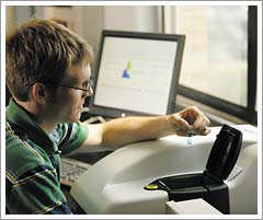
Assistant
Professor Ryan K. Roeder and Associate Professor Glen
L. Niebur, funded by the
U.S. Army Medical Research and Materiel Command, the Centers for Disease Control
and Prevention, and the National Institute of Arthritis and Musculoskeletal and
Skin Diseases program, are developing new contrast agents for micro-computed
tomography of microdamage in bone. The goal of their work is to design compounds
that, binding to a microfracture, will more accurately identify damaged tissue
on a CT scan or X-ray. These new contrast agents could help researchers develop
applications for assessing the effects of the damage to bone strength, load capabilities,
and fracture susceptibility, improving the diagnosis and prevention of fractures.
Working with Roeder and Niebur are Mark Z. Zhang, a postdoctoral researcher in
the department; graduate students Matt Landrigan and Ryan
Ross; and undergraduates
Carl Berasi and Matthew Meagher. |
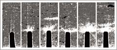 |
<< As
part of the “Contrast
Agents for Micro-computed Tomography of Microdamage in Bone” project,
University researchers employ 2-D, above, and 3-D micro-CT images
of notched, cortical bone specimens. The bright region above each
notch is stained with a heavy metal contrast agent to highlight microdamage.
Shown from left to right, the “stain” or damage increases
as the number of loading cycles is increased. The images shown here
were generated by Xiang Wang (Ph.D., AME ’05) and Huijie Leng
(Ph.D., AME ’06) in conjunction with Assistant Professor Ryan
K. Roeder and Associate Professor Glen L. Niebur. |